
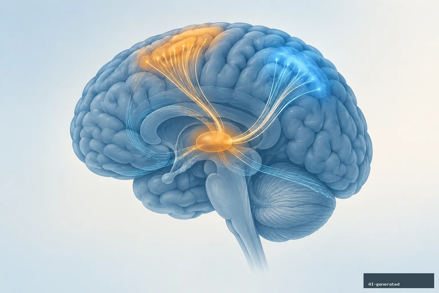
Adults assessed about two months after a concussion had lower fiber density in two right-sided brain pathways connecting the thalamus with the motor cortex and parietal lobe, according to a 2026 study. The difference appeared even though the concussion group performed similarly to an injury-control group on a series of thinking tests.
The findings point to subtle, group-level differences in white matter after mild traumatic brain injury. They do not show that the changes cause symptoms, predict recovery or indicate that an individual needs treatment.
What the study examined
Researchers compared 39 adults with mild traumatic brain injury with 28 adults who had been injured in similar accidents but had not sustained a blow to the head. Participants were recruited at two hospitals in Melbourne, Australia, after injuries occurring between 2017 and 2023.
The participants were 18 to 60 years old, with an average age of about 37 in both groups. Each person completed one MRI session and one set of cognitive tests between six and 12 weeks after the injury—about eight weeks later on average.
The concussion group met a definition of mild traumatic brain injury based on a Glasgow Coma Scale score of 13 to 15 along with brief confusion, memory loss or loss of consciousness. Two-thirds had a full score of 15.
Differences were limited to two right-sided pathways
The researchers used a method called fixel-based analysis to examine six thalamus pathways on each side of the brain. These pathways run toward areas including the prefrontal, premotor, motor, sensory, parietal and occipital cortex.
Fiber density is an estimate of how much axon material runs in a particular direction. Lower density may reflect a reduction in axons, but that interpretation is not definitive. The analysis measured fiber density, not the cross-sectional size of the pathways.
After accounting for age and sex, the concussion group had lower fiber density in:
- the right thalamus-to-motor-cortex tract, with a Bayes factor of 6.85;
- the right thalamus-to-parietal-lobe tract, with a Bayes factor of 3.71.
The researchers described these results as moderate evidence. Evidence for a lower fiber density in the matching left thalamus-to-motor-cortex tract was weak, with a Bayes factor of 1.28.
There was no clear difference in the other nine thalamus pathways. A whole-brain analysis also found no differences that remained after correction for multiple comparisons.
Normal test scores do not rule out subtle findings
The two groups scored alike on all seven measures covering attention, processing speed, memory and mental control. This means the MRI differences were not accompanied by measurable group differences on the cognitive tests used in this study.
Some exploratory analyses linked fiber density with test performance, but the patterns were not straightforward. For example, higher fiber density in the left thalamus-to-premotor pathway was associated with slower processing speed in the concussion group and faster processing speed in the control group. Among controls, higher fiber density in several right-sided pathways was associated with lower digit-span scores; that pattern was not seen after concussion.
Because the researchers ran many exploratory comparisons across 12 pathways and seven tests, these associations require further study. They do not establish that fiber density changes affect thinking or explain symptoms.
Why the results need cautious interpretation
The study provides only a snapshot. Participants had one scan, and the researchers did not have scans from before the injuries. As a result, the study could not determine whether the differences were caused by the concussion, existed beforehand or later faded or persisted.
The research team suggested that fiber density might rise soon after concussion because of swelling and fall later if axons degenerate. The current findings at about two months fit that proposed later phase, but the study did not demonstrate this sequence.
The participants were hospitalized adults injured mostly in road accidents and falls. The findings may not apply to athletes, veterans, people injured in assaults or people with repeated concussions, who were excluded.
What the findings mean for readers
Advanced MRI measures such as fiber density can reveal patterns that are not captured by standard cognitive testing, but fixel-based analysis is not presented as a clinical diagnostic test. The study also does not establish a treatment response or a cause for any individual’s symptoms.
For now, the main value of the research is in understanding how mild traumatic brain injury may affect specific thalamus connections over time. Larger studies with pre-injury scans and repeated follow-up imaging will be needed to determine whether these differences are injury-related and whether they persist.
AI tools were used to assist with the preparation of this article.
Leave a Reply