
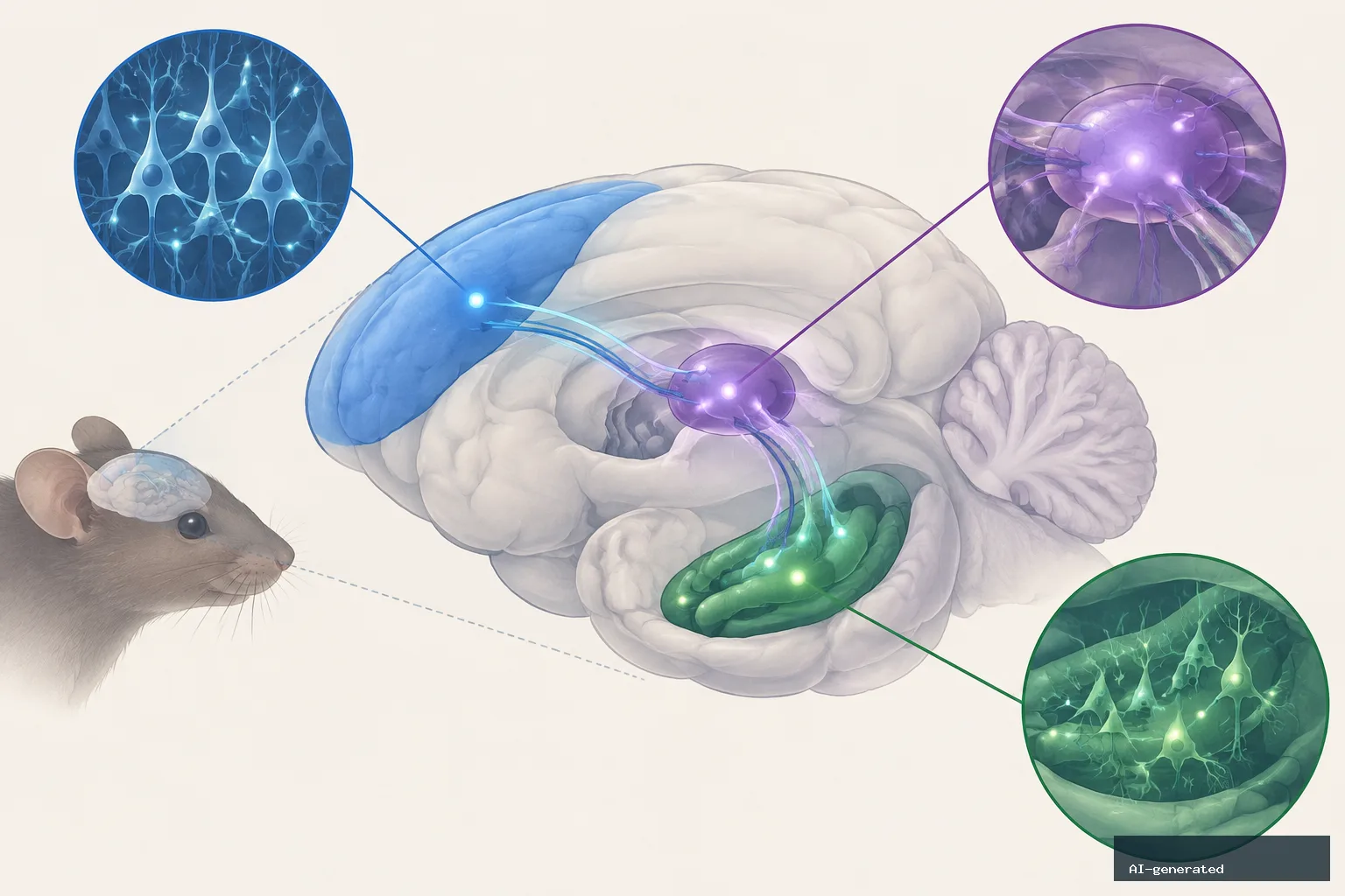
A brain pathway linking the prefrontal cortex, a midline thalamic region and the hippocampus appears to be necessary for ketamine’s antidepressant-like effects in stressed mice, according to a 2026 study published in Nature Communications.
The research focused on the nucleus reuniens, a relay in the midline thalamus that connects the prefrontal cortex with the hippocampus. When researchers temporarily switched off the relay—or either end of the pathway through it—ketamine no longer reduced immobility in a swim test used in the study.
The findings identify a circuit required for the observed response in mice. They do not show that the same pathway is required for ketamine to work in people, nor do they establish where in the brain the drug acts.
What the researchers tested
The study used mice exposed to a five-day stress procedure. Afterward, the animals showed several stress-related behavioral changes, including less interest in sugar water, reduced exploration and faster loss of active struggling in swim and tail-suspension tests.
Researchers gave some stressed mice a single 10 milligrams-per-kilogram dose of ketamine. With the circuit intact, the drug reduced immobility in the swim test. That effect was not observed when the nucleus reuniens was silenced.
Silencing prefrontal fibers entering the nucleus reuniens, or fibers leaving the reuniens for the ventral hippocampus, also removed the observed behavioral response. The behavioral groups were small, ranging from four to 17 mice.
The researchers used chemogenetics to control selected neurons and projections. In this system, hM3Dq served as an activating switch, hM4Di as a silencing switch and mCherry as a control virus.
Links to hippocampal plasticity
The team also examined long-term potentiation, or LTP, a cellular measure of the ability of connections between neurons to strengthen. LTP was absent in hippocampal slices from stressed mice compared with slices from unstressed mice.
Ketamine restored LTP in the slices. Blocking the prefrontal-to-reuniens connection prevented that restoration, tying the behavioral finding to a change in hippocampal plasticity.
The researchers then activated excitatory neurons in the infralimbic cortex—the mouse region described as corresponding to part of the human medial prefrontal cortex—overnight. This reduced immobility in both swim and tail-suspension tests, increased sugar-water preference and improved novel-object and social exploration.
The overnight activation also restored LTP. By contrast, a single acute activation did not change the behavioral measures or restore LTP. Activating the prelimbic cortex, another prefrontal region, did not change immobility or sugar-water preference.
General movement did not appear to explain the results: open-field distance traveled and ordinary home-cage exploration were unchanged after overnight infralimbic activation.
A selective connection to the ventral hippocampus
Additional analyses pointed to the ventral rather than dorsal hippocampus. Markers of incoming thalamic connections increased in the ventral hippocampus, while the change in the dorsal hippocampus did not meet the study’s statistical threshold.
Several plasticity-related genes were activated in the ventral hippocampus, with no such activation detected in the dorsal hippocampus. Markers of local and inhibitory connections did not change.
Silencing the nucleus reuniens canceled the behavioral and plasticity effects of prefrontal activation. Silencing the reuniens by itself did not change immobility, suggesting that the relay’s involvement depended on the wider circuit rather than simply on its activity in isolation.
Why the human connection remains uncertain
Ketamine acts on receptors throughout the brain, including directly in the hippocampus. The study therefore does not establish that the nucleus reuniens is where ketamine acts. The authors proposed two possibilities: the drug may act locally while requiring an engaged prefrontal–thalamus–hippocampus loop, or it may act at several points and require the loop to carry the effect forward.
Earlier rat research cited by the source also implicated the ventral hippocampus and its connection with the prefrontal cortex. Separately, deep-brain stimulation for treatment-resistant depression targets the subcallosal cingulate cortex, described as corresponding to the mouse infralimbic cortex.
A white-matter study of 16 patients found that people who responded to stimulation shared bilateral connections from the stimulated area to medial frontal cortex, cingulate cortex and subcortical nuclei. That finding was anatomical overlap, however, not confirmation that the mouse mechanism operates in people.
No person has been tested to determine whether the midline thalamus is required for ketamine’s effects. The source describes the practical value of the mouse work as a possible target for future research, not as a change to current ketamine or brain-stimulation use.
What the study adds
The results suggest that antidepressant-like effects in this mouse stress model depend on coordinated communication across several brain regions. They also distinguish between a circuit that may be necessary for an effect and the site where a drug initially acts.
Researchers were blinded to treatment groups until data collection, randomly assigned the mice and included equal numbers of males and females. The study was not powered to detect sex differences, and its exploratory analysis found none.
Three authors reported lecture fees or advisory honoraria from Johnson & Johnson, which makes esketamine.
Key takeaways
- In stressed mice, ketamine reduced swim-test immobility only when the nucleus reuniens and its prefrontal-to-hippocampus connections remained active.
- Silencing either the prefrontal input to the nucleus reuniens or its projection to the ventral hippocampus removed the observed response.
- Ketamine restored hippocampal long-term potentiation in stressed mice, but blocking the prefrontal-to-reuniens connection prevented that restoration.
- Overnight activation of the infralimbic cortex reproduced several behavioral and plasticity findings, whereas acute activation did not do so in the reported tests.
What this means
The study is relevant because it identifies a specific brain network involved in ketamine-related effects in a mouse stress model. Its findings may help guide future research into brain circuits, but they do not establish a human treatment mechanism or change how ketamine or brain stimulation is used.
What the evidence cannot tell us
- The ketamine circuit experiments were conducted in mice, not people.
- The stress-related behaviors in mice are not depression itself.
- The study does not establish that the nucleus reuniens is where ketamine acts.
- No human study has tested whether the midline thalamus is required for ketamine to work, and the human stimulation comparison was anatomical overlap rather than confirmation of the mouse mechanism.
AI tools were used to assist with the preparation of this article under our editorial standards.
Leave a Reply